Home Cytometry History Individual Histories Howard Shapiro

Howard Shapiro

I literally grew up with cytometry; I don’t know when I first made the acquaintance of a microscope, but it had to have been before I was six years old. My mother, who had interrupted her graduate study in biology to have two kids, worked at Brooklyn College; we lived a very short distance from the school and, especially during World War II and the years immediately after, before I started school myself, I was often taken to the lab where a maternal eye could be kept on me. The smell of cooking agar mingles with that of chicken soup in my childhood memories.
Arthur Pollister was a great analytical cytologist at Columbia University; he and Len Ornstein wrote an extensive chapter on absorption measurements in Mellors’s book, “Analytical Cytology.” Arthur’s wife, Priscilla, taught at Brooklyn, and my parents were friendly with the Pollisters, but I don’t know whether I absorbed any cytometry while being bounced on Arthur’s knee as a toddler. Around 1950, Brooklyn College acquired an electron microscope, and my mother became the operator; she also went back to graduate school (at New York University), switching her field from microbiology to histology and cytology. She was working on her thesis when I was in High School, and I remember her teaching me how to do some histochemical stains, the Feulgen included, so I could help her out in the lab after school. One of the professors on her thesis committee, M. J. Kopac, had built a video microscope which I remember seeing some time in the early 1950s. Remember, nothing was computerized then, but you could take a photo of the TV screen. Kopac’s gadget also had the capability of feeding a single raster line into an oscilloscope, producing a trace of the transmission (or extinction) at various points along the line, which could also be photographed. By the time I was doing science projects in high school, I had started reading Caspersson’s “Cell Growth and Cell Function” and Jean Brachet’s “Biochemical Cytology.” Brachet had resurrected the methyl green-pyronin Y stain, originally described in the 1880s, as a semiquantitative stain for DNA and RNA, and, remembering that many years later when I encountered papers published in the late 1970s that showed methyl green and Hoechst dyes bound similarly to A-T pairs in DNA led me to the Hoechst/pyronin Y stain for DNA and RNA. History is often the best teacher; my mother would probably have been an even better one but she unfortunately succumbed to lymphoma in 1963, just six years after receiving her Ph.D., and I was pretty much stuck with history from that point on.
While I was in medical school in the 1960s, I worked with computers, spending some time on making metabolic flux models of cells (I published on this in the 1960s; other people reinvented the concept in the mid-1970s and it now seems to be a fairly big deal in systems biology) and some on analysis of EKGs. We recorded EKGs on analog equipment at Bellevue Hospital, in Manhattan, and then carried the 9-track tapes by subway to NYU’s engineering research facility in the Bronx, where they were laboriously digitized and crunched by a Control Data mainframe somewhat larger than an IBM 7094. There were few enough people working with computers in biology and medicine then so that there would typically only be one big meeting on the subject every year or so, and it was at some of those meetings that I first ran across Mort Mendelsohn, Brian Mayall, and Judy Prewitt, who were then trying to automate white cell identification using the CYDAC flying spot scanner at the University of Pennsylvania. Being able to program and use computers got me a research job at NIH, specifically at the National Cancer Institute, where the objective was to build a system that would identify leukemic blasts in emulsion-coated marrow slides and determine cell kinetics by autoradiographic grain counting. NCI sent me to a New York Academy of Sciences Meeting on computers in biomedical imaging in June, 1967, just before I left New York (where I had been a surgical resident) for Bethesda. I met Lou Kamentsky and Mike Melamed at that meeting, and also Ken Preston, then at Perkin-Elmer, who had built a microscope scanner used in some pilot studies for NCI. Also aiding NCI in the autoradiograph project were Lew Lipkin, a pathologist from the National Institute of Neurologic Diseases (NINDS), and Russell Kirsch, a computer scientist from the then National Bureau of Standards (NBS, now NIST), who had, in 1957, been the first person to digitize a photograph. An old friend of mine from high school, Phil Stein (now, unfortunately, no longer with us) was working at NBS at the time, hooking up computers to various experimental apparatus, and he told me he would like nothing better than to work with Russell Kirsch. When I arrived at NCI, I suggested two things to the powers that were. One was that we pool resources with NBS and the NINDS, ransom the scanner from Perkin-Elmer and have it rebuilt with appropriately rugged and precise microscope hardware attached at NBS and then installed at NCI. That was done. The second suggestion was that NCI put money into flow cytometry, preferably in Bethesda as well as elsewhere. We did put money into Los Alamos (and, later, Livermore), and some into Kamentsky’s early work, but the first flow cytometers at NIH, built at Los Alamos, didn’t arrive until I after left to do clinical work in 1970. I did, however, get to keep up with the field thanks to meetings I attended in the late 1960s, at which I met Bob Leif, Mack Fulwyler, Ted Young, and some other members of the old guard. I missed the first two Engineering Foundation Conferences on Automated Cytology because I was doing clinical things at the time.
By 1972, I was working in medical instrumentation at G. D. Searle &. Co., a Chicago-area drug firm (now engulfed into Pfizer) with interests in diagnostics and medical devices. Their Nuclear-Chicago subsidiary, which made scintillation counters and gamma cameras, had the dubious distinction of being offered the CAT scanner and turning it down. One of my jobs was to evaluate instrument proposals from outside Searle, and, in early 1973, we were visited by Myron Block, Tomas Hirschfeld, and Bob Schildkraut, of Block Engineering, then in Cambridge, Massachusetts, who proposed to build a couple of flow cytometry-based diagnostic instruments. The first would be a hematology counter, using five illuminating beams and measuring eight parameters, including four wavelengths of fluorescence; it would process 50,000 cells/second, producing a 1,000-cell white cell differential count on the fly in less than two minutes. The second would be a slow-flow system with near single-molecule sensitivity, intended to detect single hepatitis B virus particles in blood samples. Block was already producing the first commercial Fourier Transform Infrared Spectrophotometer, with a built-in Data General minicomputer, and, thanks to my apprenticeship at NBS while we got the NCI-NINDS-NBS scanner together, I could appreciate that they really knew what they were doing as far as the instrumentation went, and enthusiastically recommended that Searle fund the projects. Once the prototypes got built, it became clear that the otherwise stellar Block crew didn’t know as much about cells as I had thought, and I ended up having to commute to Boston weekly and, in 1975, to move there, rather than monitoring the project by phone from Illinois with an occasional Boston trip. If my mother had been alive, I probably could have gotten the projects onto the right track with a few phone calls to her; not having a reliable medium on hand, I had to dig into the literature. Medline only went back to 1965, so this meant spending a lot of time in the bowels of the Harvard Medical library.
The Third Engineering Foundation Conference on Automated Cytology was held at Asilomar in December, 1973; I found out about it only a few months beforehand, but was able to wheedle Mort Mendelsohn, who had by that time moved to Livermore and was chairing the meeting, to make room for me and some of the Block people. Myron Block was playing his cards close to the chest at that time, so we couldn’t tell anybody what we were doing, but we did get to mingle with the Herzenbergs and their group and with people from Los Alamos and Livermore. Tom and Donna Jovin and some folks from Europe also attended that meeting, but, if I recall correctly, Wolfgang Goehde and his colleagues did not; the American and European flow people didn’t get fully integrated until some of the European meetings a few years later.
By 1975, when Myron allowed us to talk about the Block systems at the next Asilomar meeting, it became obvious that, although they didn’t sort, they were pretty far ahead of the rest of the field in terms of their measurement and data analysis capabilities (too far, as it turned out, to be usable in clinical environments), and I started to try to get the technology transferred to one or another of Boston’s well-known medical research institutions. In 1977, I took over the Cytokinetics lab at the then Sidney Farber, now Dana-Farber Cancer Institute on a part-time basis, Awtar Krishan having left for Miami, and started to cobble the first of the Cytomutts together using parts from Block prototypes, a Bio/Physics Systems Cytofluorograf, and some apparatus built in Caspersson’s lab in Stockholm for his collaborative work with Sidney Farber, George Foley, and Ed Modest on chromosome banding. By the mid-1980s, I had set up flow labs at a few Harvard-associated institutions, helped a couple of groups at MIT, including Penny Chisholm’s, build flow cytometers, and consulted with various industrial organizations, and it became easier to get research funding through my company than to deal with the various academic administrations. I had also become known for presenting work in song, verse, and on T-shirts and mobiles.


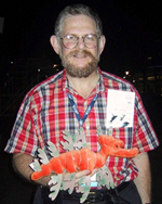
Howard playing Crème de la Crème for Nobel Laureate César Milstein and SAC XII attendees in Cambridge, England, 1987
To listen to some of Howard’s songs – click on a title
Being able to work with multibeam computerized apparatus years before others did was good because we could do experiments that others could not, but bad because it was not always easy to explain what we were doing. In 1983, I published a paper called “Multistation Multiparameter Flow Cytometry: A Critical Review and Rationale,” in which I tried to make the cytometry community aware of the benefits; this was originally a shared instrument grant application that didn’t, at first, get funded, and it metamorphosed into “Building and Using Flow Cytometers: The Cytomutt Breeder’s and Trainer’s Manual.” Bob Schildkraut (of Block) and I had written a chapter in the original 3M book on “Flow Cytometry and Sorting,” published in 1980, of which Wiley sold 1,000 copies; by 1983, Alan Liss, who was publishing Cytometry, was interested in publishing a flow cytometry text, and I agreed to turn “Building and Using…” into “Practical Flow Cytometry.” At the time, I figured I knew a lot about flow cytometry, and that it wouldn’t be right to run off and do something else without writing a reasonable amount of it down. I was giving serious thought to moving into molecular biology or experimental oncology. It never happened; once the book came out, it became impossible for me to escape from cytometry, and I’d have to say that, even after more than a generation, it hasn’t been all that easy for me to move away from flow cytometry back toward imaging.
In retrospect, I am very lucky to have been able to maintain a respected position in the field without having to run a large lab on a large budget. I am not under pressure to process large numbers of specimens either for myself or for others, and I have more time to think than just about anybody else I know. I never intended to retire (which I probably would have had to do at some point in a more formal environment), but I suspect that either my oncogenes or some other as yet unclarified genetic peculiarities may catch up with me before I become entirely irrelevant to cytometry. I’ll just try to hang on as long as I can.
