
Home Cytometry History Individual Histories Harry A. Crissman

The Serendipitous Journey to Los Alamos and Flow Cytometry
by Harry A. Crissman
When I was 23 years old, and most of my high school classmates had already earned their bachelor or advanced degrees, I was drafted into the US Army where I served for two years in the 101st Airborne Division. At that point, I had not a clue of what career path to follow after my Army discharge. Fortunately, I decided to return to college where I received my BS degree in teaching from Lock Haven University Pennsylvania, a small college in my home town. Still without a clear direction, I worked for a few years in a family meat packing business. Eventually, at age 30, I took a two-year position teaching high school biology and chemistry in Ovid, NY before attending Pennsylvania State University, where I obtained a MS and Ph. D. Then, in August 1971, as luck would have it, I accepted a postdoc position at Los Alamos National Laboratory (LANL) – truly the right place at the right time. My initial assignment at Los Alamos was to develop fluorochrome staining techniques that would improve existing resolution of DNA content in single cells by flow cytometry. Increased resolution in DNA content histograms (i.e., decreased CVs) allowed for the eventual development and verification of computer-fit, cell-cycle frequency analysis. My Ph.D thesis involved DNA, RNA and protein staining of tissues sections of liver with non-fluorescent, stochiometric, absorption dyes and cell-by-cell analysis by microspectrophotometry, a slow microscopic process yielding analysis of about 250 cells per day. Flow cytometry (FCM) was an entirely new concept for me, and, although, prior to going to Los Alamos, I had received copies of the Fulwyler cell sorting paper (Science 1965) and the Van Dilla et al DNA analysis paper (Science 1969), I had no idea how this new FCM technology could perform analysis at rates of thousands of cells per second. As time passed I began to realize and truly appreciate how much time, dedication and ingenuity the Los Alamos group had put into the effort to develop such a precision instrument that would be available to me upon my arrival. After my first session on the FCM, I remember Marv Van Dilla remarking to me, “Do you realize that you analyzed more cells this afternoon than you analyzed during all your four years at Penn State.” It was true!
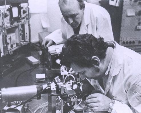
Harry Crissman (foreground) and Marv van Dilla preparing to analyze DNA content in stained cells in the single parameter flow microfluorometer (FMF) at Los Alamos National Laborotory in the early 1970s. The term FMF was used to describe the instrument at that time, before the name was later designated to be a flow cytometer (FCM).
During my 33-year career at Los Alamos, I had the gifted opportunity to work with a unique group of dedicated scientists – Van Dilla, Fulwyler, Steinkamp, Martin, Tobey, Mullaney, Cram, Dean, Jett, Kraemer and so many others, all of whom took me under their wings and taught me the basic arts of cell cycle analysis and flow cytometry. I am truly indebted forever to the generosity of these individuals for taking the time and having the patience to help me to develop a career that I never could have even dreamed of achieving during that ever deviating journey that finally led to Los Alamos. Early on we developed new Feulgen staining methods for cell cycle analysis methods and later introduced DNA-propidium iodide (PI) labeling, initially with a gratis PI sample obtained from Bruce Hudson, Jerome Vinograd and collegues who had just recently synthesized the dye for improved detection and isolation of closed circular DNA by dye-buoyant density procedure.
Bob Tobey and I published the first paper on the use of FCM for detailed analysis of the effects of chemotherapeutic drugs on cell cycle progression, and, a year later, Bob Tobey and I developed a rapid method for specifically staining DNA with mithramycin, a compound that was being used to treat testicular cancer at the time. The technique led to the publication in Science, “Cell Cycle Analysis In Twenty Minutes.” The method is not often used today, but following the publication, and a large number of citations, cell biologists and biochemists began to appreciate flow cytometry as a useful methodology and not just a technical novelty. I still remember receiving a surprising phone call from Robert Holley, the 1968 Nobel Prize winner, requesting a vial of mithramycin.
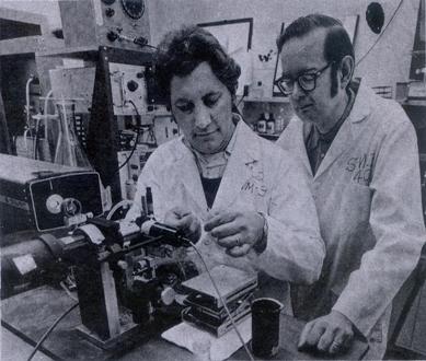
Harry Crissman (left) and Bob Tobey preparing to analyze the DNA content in stained samples of cells in the Los Alamos flow microfluorometer in the early 1970s. The cell population had been previously treated with cancer chemotheraputic agents in some of the first (FMF) studies designed to determine the effect of the agents on cell cycle progression.
Later we, including Darzynkiewicz and Steinkamp, were first to demonstrate simultaneous FCM analysis, including ratio analysis of DNA, RNA and protein (Science, publication) and later developed, with Steinkamp, a FCM method for lifetime analysis of DNA-bound fluorochromes for detecting changes in chromatin structure, for which one of two Research and Development 100 awards was presented. I was very fortunate to have worked with John Steinkamp, since it was his ingenious development of multiparameter instrumentation that made possible so many of the biological techniques that I was able to develop and utilize. In a PNAS publication we, including Tobey, Gadbois and Bradbury, showed clear cut differences in kinase-mediated regulation of cell cycle progression in normal and transformed cell, an important factor that is currently incorporated into the development of new cancer chemotherapeutic drugs.
Serving as president of ISAC was truly an honor and a wonderful segment of my career. To be elected by the membership and to follow in the path of so many others, presidents, councilors and so many members who had generously volunteered their time to make our society one of the very best was a dream come true. As a charter member, I had the opportunity to follow the membership growth in ISAC from several hundred to several thousand and to watch the development and success of the journals, Cytomety, parts A and B. But ISAC is more than a regimented society; it is a band of scientists working together in the development of unique techniques for cellular analysis for use in basic research and medical science. As in the past, ISAC will continue to lead the way to new discoveries. I am proud to be a member of ISAC.

- Harry A. Crissman, Past-President of ISAC 2000-2006