The First Flow Cytometry Reagents
For Intracellular Enzyme Analysis
| |
CellProbeÖ Reagents The First Flow Cytometry Reagents For Intracellular Enzyme Analysis |
Flow Cytoenzymology: The Inside Story
In Flow Cytoenzymology, scientists utilize flow cytometry and fluorogenic substrates to measure intracellular enzyme activity. For almost two decades, researchers have recognized the potential contribution of this field to the understanding of cell function. However, only a few dedicated researchers1-3 have been able to devise successful methodologies. Many technical challenges -- such as cell permeabilization, enzyme cross-reactivity, the need to maintain cell viability, and the sensitive nature of a kinetic assay -- have kept this field largely untapped.
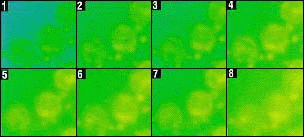 |
| Pseudo-color display of fluorescence intensity in sequential time intervals shows how the fluorescent product from cleaved substrates accumulates in cells, reaching maximum fluorescence emission in a matter of minutes. This experiment was conducted with leukocytes stained with CellProbe AĽAminopeptidase M and analyzed on an image analysis system. (Courtesy of F. Lucas, Coulter Corporation). |
Introducing: The CellProbe Reagent Line
Coulter introduces an innovative new line of reagents that makes Flow Cytoenzymology a reality. The CellProbe reagent line offers a selection of synthetic substrates designed specifically for flow cytometry. The unique reagent formulations (patents pending) allow the substrates to pass through the cell membrane unhindered -- without the need of a permeabilization step. Enzyme specificity and activity are enhanced through a carefully selected combination of cofactors and inhibitors. Inside the cell, enzymatic hydrolysis of the substrate frees a fluorescent dye (either fluorescein or rhodamine 110) which accumulates in the cell as a function of the concentration or activity of the enzyme. The resulting fluorescence is easily analyzed by flow cytometry, as well as other methods for detecting cellular fluorescence.
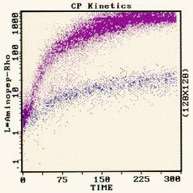 |
| Two parameter kinetic display of CellProbe LĽAminopeptidase activity vs. TIME shows the fluorescence change over time, in seconds. Enzyme activity in normal cells (blue) increases only slightly. Leukemic cells (purple) increase in fluorescence intensity nearly a thousand-fold in the course of 5 minutes. (Courtesy of J. Hoffman, G. Jaffe, Coulter Corporation). |
| |
Research Opportunities Whole Blood To Histograms In Minutes The Leader In Cell Analysis Reagent List Slide Presentation |
*For Research Use Only. Not For Use In Diagnostic Procedures.
1Dolbeare, FA and Smith, RE, 1977. Flow cytometric measurement of peptidases with use of 5-nitrosalicylaldehyde and 4-methoxy-ß-naphthylamine derivatives. Clin Chem 23:1485-1491.© Coulter International Corporation, 1996. All rights reserved. Copyrights and Trademarks
 |
 |
 |
 |