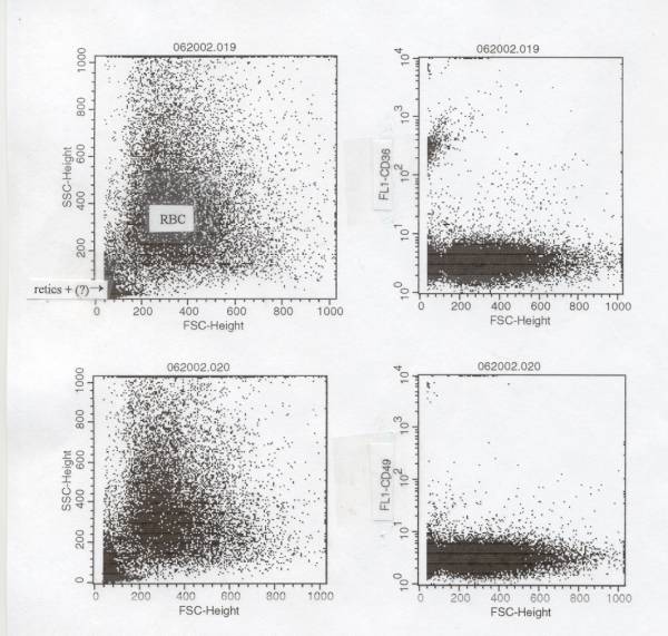
Hi,

Can someone working with human reticulocytes tell me if it's possible to get
both retics and RBC on a forward by right angle light scatter dot plot?  In
case it's relevant, the blood samples will be from patients with sickle cell
disease.

We are trying to gate RBC and retics by light scatter and then use a number
of antibodies (CD36, CD49, and annexinV) to characterize these cells so we
would prefer not to use a fluorescent stain for detecting the cells but
rather to save the fluorescence PMTs for antibody/marker fluorescence.

Also, in case it's relevant, we're using a 3-color FACSVantage SE for these
analyses.

Finally, if someone knows of a publication is which is illustrated (by
scatter dot plot) the detection of retics by light scatter, will you please
give us that reference.

The attachment shows the light scatter and fluorescence of one such sickle
cell disease sample stained with CD36 or CD49d.  You can see the retics in
the bottom left corner (they stain with CD36 but not with CD49d).  What we
need, however, is to be able to get all of the retics on the dot plot.
Increasing the FSC gain (for these samples it was at 8 using the 488 line at
200 mW) doesn't seem to help much.  I guess the question is, should we be
able to, if we did something differently and/or better?

Thanks very much for any advice/references.

Ray Hester
Univ. of South Alabama
rhester@jaguar1.usouthal.edu
This archive was generated by hypermail 2b29 : Sun Jan 05 2003 - 19:26:14 EST