Using the best of the current manual (reference method) and the best of the current flow cytometric methods, Coulter applies 3-D VCS Technology to the study of peripheral Red Blood Cells. Available as an inexpensive upgrade to existing COULTER® STKS, MAXM and MAXM A/L Hematology Systems, the Coulter VCS Reticulocyte Method gives the routine hematology laboratory a simple, fast, accurate and cost effective Reticulocyte count.
A blood sample is first incubated with the supra vital stain New Methylene Blue (NMB) for 5 minutes. The dye precipitates any residual RNA within the erythrocytes. A small portion of the stained blood sample is then diluted with a hypotonic acid solution that will clear the erythrocyte of hemoglobin, but preserve the stained RNA within the cell.

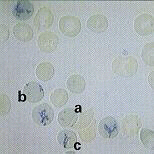
Blood Stained with NMB
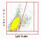
Reticulocytes in various stages of maturity are pictured at the left. The younger cells have more residual RNA and therefore stain more heavily. When illuminated by a HeNe LASER, these cells scatter the most light. Older Reticulocytes have little residual RNA and stain less intensely. However, because of the clearing step, they are easily separated from mature erythrocytes by light scatter.
The diluted sample is aspirated into the STKS or MAXM and analyzed in the VCS Flow Cytometer. This analysis provides simultaneous measurement of a cell’s volume, internal structure, granularity and cell surface characteristics.

Using the powerful 3-D VCS technology, over 30,000 cells are rapidly analyzed. Reticulocytes are separated from the mature RBC’s and expressed as a percent. If run as a profile that includes an RBC count, the results will also be expressed in absolute number.
For more information or to obtain this upgrade for your current Coulter STKS, MAXM or MAXM A/L, contact your local Coulter company.


