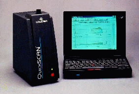
Coulter Nav - Other Products

COULTER QuickSCAN

Quickscan
COULTERTM QuickSCANTM
A NEW CONCEPT IN STABILITY ANALYSIS OF CONCENTRATED
COLLOIDAL DISPERSIONS (EMULSIONS, SUSPENSION, FOAMS, GELS)

Principle

The design principle of the Coulter QuickScan is the association of scanning and backscattering/transmission detectors.

The optical system is designed around a new concept of a reading head which combines transmission and backscattering. It works in the near infrared (= 850 nm) where there is little absorbance of light. It is sensitive to two kinds of parameters:

1. Particle concentration: Clear or slight turbid systems are analyzed in transmission (<0.1 % of particle concentration) while turbid to very opaque mixtures are analyzed using backscattering (>0.1 % of particle concentration). The high level of the backscattering signal for low particle concentration is due to the second reflexion of the cell glass. This value increases with the clearness of the sample and follows the evolution of the transmission. In fact, it does not need interpretation. Backscattering is also able to measure very concentrated mixtures (50% particle amount for low refractive index mixtures like emulsions) and black systems.

2. Particle size: It is known that backscattering maximum is obtained with a particle size between 0.5 and 1 µm. Backscattering intensity decreases when particle size is above or below this value. This is in accordance with the Mie theory which states that the light scattering is at a maximum when particle size approximates to the wavelength used (850 nm for the QuickScan).

The measurement cells are usually filled with 5 ml sample. The sample stays in the cell during the entire aging process.

Applications

In comparison to other analytical instruments, the QuickScan presents an advantage of working with opaque and concentrated mixtures without the need of previous dilution. This performance makes it particularly useful to analyze the stability of emulsions, suspensions, gels and foams. The mixtures are particularly used in Cosmetics and in Pharmaceuticals as well as in other industries like agro-food, agrochemical, paints, detergents and chemistry in general.

The naked eye cannot detect or quantify the two kinds of destabilization of colloidal dispersions at an early stage.

1. Particle migration: This reversible phenomenon is due to the difference in density of the dispersing and the dispersed medium. It this case, the particle concentration change commences at the top and bottom of the sample only (creaming, sedimentation).

2. Particle a aggregate size variation: This irreversible process is undetectable by the naked eye at an early stage (coalescence, flocculation).

The QuickScan detects particle size and concentration variations in the mixture by scanning the whole height of the sample in transmission and in backscattering. It displays the sample profile as a macroscopic graph.

Thus, transmission and backscattering profiles are flat on the whole height of the sample for a homogeneous mixture. If the mixture remains stable as a function of time, these profiles do not change. When particles migrate to the top or bottom of the sample (creaming, sedimentation), transmission and backscattering levels vary proportionally to the particle concentration in each area. On the other hand, if the particle or aggregate size increase as a function of time (coalescence, flocculation), transmission and backscattering levels change within the whole height of the sample.

Controls, Standards and Accessories


Home Contents Previous Next
© Coulter International Corporation, 1996. All rights reserved. Copyrights and Trademarks

Home Page Table of Contents Sponsors Web Sites
CD ROM Vol 2 was produced by staff at the Purdue University Cytometry Laboratories and distributed free of charge as an educational service to the cytometry community. If you have any comments please direct them to Dr. J. Paul Robinson, Professor & Director, PUCL, Purdue University, West Lafayette, IN 47907. Phone:(317) 494-0757; FAX (317) 494-0517; Web http://www.cyto.purdue.edu EMAIL robinson@flowcyt.cyto.purdue.edu