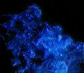
Out-of-focus haze removed using VayTek's deconvolution software. |
What Is Deconvolution?
How Does Digital Imaging
Relate to Microscopy?
Frequently Asked Questions about
HazeBuster/MicroTome (HB/MT)
and Digital Imaging Systems
HazeBuster is VayTek's most affordable package
MicroTome is VayTek's best deconvolution software, with
the most features |
- What is HB/MT?
- What is a confocal microscope?
- How does HB/MT work?
- What is the value of a confocal image?
- What are the advantages of HB/MT
over a confocal microscope?
- What are the limitations of
HB/MT?
- What is a deconvolution algorithm?
- How do you adjust the haze removal
with HB/MT?
- Will the deconvolution approach
replace the confocal microscope?
- How does the nearest neighbor algorithm
work?
- What is a Point Spread Function (PSF)?
- Is a theoretical PSF accurate enough
to produce high quality images?
- How do I know that what I see in the deconvolved
image is real?
- How fast can HB/MT deconvolve an image?
- Is HB/MT easy to use?
- What are the data acquisition issues?
- What image formats are read by HB/MT?
- Is there technical support for this
product?
- How can I visualize my data?
- Can I get a hard copy print out of my images?
1. What is HB/MT?
HB/MT software uses the nearest neighbor algorithm to mathematically
calculate and remove out-of-focus haze from microscope images. It is designed
to replace or supplement pinhole-based confocal microscopes.
2. What is a confocal microscope?
Minski (1961) was the first to propose the technique of confocal microscopy
used by laser scanning confocal microscopes. The principle is quite simple
and is illustrated in the light paths in Figure 1.

The image seen through a microscope includes the in-focus portion and
the out-of-focus portion above and below the plane of focus. The smear or
blur produced by the out-of-focus planes is a natural consequence of the
optics of the microscope.
Confocal microscopy removes out-of-focus haze by passing the light through
one or more small apertures, leaving only a thin, highly focused plane.
The light from this focused plane can be digitized and stored on a computer.
The distance between the specimen and the microscope objective is then
changed producing a new focal plane. The new focal plane is digitized and
stored. After a series of planes has been collected, individual slices can
be examined or the whole specimen can be digitally reconstructed by a computer
as a three-dimensional volume.
A confocal microscope consists of a standard microscope with a number
of complex attachments to direct and process the beam of light. Most confocal
microscopes use an intense laser light to scan the specimen. This intense
light source is needed to compensate for the light loss which occurs as
the light passes through small apertures.
3. How does HB/MT work?
HB/MT does in software what the confocal microscope does by virtue of
hardware (i.e. the pinhole). Both systems use image processing but HB/MT
is more flexible.
HB/MT, as illustrated in Figure 2, uses a standard white-light microscope
and requires no special attachments. A video camera captures and digitizes
the images from the microscope, which are then stored by a computer. Image
enhancement algorithms are used to deconvolve the image, i.e. remove the
blur or haze contributed by the out-of-focus image planes. The algorithms
used by HB/MT have the same function as the apertures in the laser scanning
confocal microscopes - removing the out-of-focus portion of the image.

You can transform your standard microscope and your computer by simply
adding VayTek's HB/MT package.
4. What is the value of a confocal image?
A confocal image has the out-of-focus haze removed. This can theoretically
increase image resolution. The increase in resolution, by as much as 1.4
times (Brackenhoff, 1989), results in improved measurements (Yelamarty,
1990) and visualization. In addition, the optical sectioning is non-invasive
and can be performed on living specimens. Also, it is possible to acquire
images with multiple wavelengths of light and merge the results for greater
information.
Besides increasing the resolution of the image, the deconvolved slices
can be stacked to produce a three-dimensional representation of the specimen.
Visualization of a three-dimensional data set can lead to new insights.
5. What are the advantages of
HB/MT over a confocal microscope?
There are a number of advantages in using HB/MT. First,
it costs less than a laser scanning confocal microscope because the microscope
you currently have can be used with the digital deconvolution approach.
New optical equipment is not required. Prices for laser scanning confocal
microscopes typically range between $75,000 and $300,000. The price of HB/MT
is a fraction of this cost.
Depending upon components already in place, you may need to add a framegrabber,
camera, and stepper motor in addition to HB/MT. For a complete description
of products, turnkey systems
and current pricng please refer to VayTek's Product
Guide or call directly (515) 472-2227).
Second, the high intensity laser light, required by
the LSM's, can harm living specimens. The digital approach is less harmful
to living material since it typically uses a small fraction of the light
used by the laser scanning microscopes.
Third, many fluorescence preparations bleach easily,
even with standard light sources. These dyes cannot be used with laser scanning
confocal microscopes. Even robust preparations can fade after many scans
producing a brightness gradient along the vertical axis. HB/MT will result
in less photobleaching.
Fourth, HB/MT is more flexible. When using a laser scanning
confocal microscope, the amount of haze removed is set by the aperture size
and thus cannot be adjusted after the image is captured. HB/MT, on the other
hand, allows the user to set the amount of haze to be removed as a part
of the deconvolution process after the image has been captured. Thus, the
HB/MT user can explore the same data set multiple times with different degrees
of haze removal.
And Fifth, data acquisition for HB/MT can be faster
than confocal microscopes. Video cameras, used by HB/MT, can average or
integrate several slices per second. Including the time to move the stage,
three images can be captured in about 3 seconds. Each image is then deconvolved,
requiring no more than four seconds (MicroTome version) per 512 x 512 slice.
Confocal microscopes can be slower. Each slice is often scanned and integrated
multiple times with reduced laser power in an attempt to attenuate photobleaching
effects.
6. What are the limitations of
HB/MT?
The principal limitation of the digital deconvolution approach has been
the amount of computer time required to deconvolve a single slice. Until
now, a single slice could require several minutes to deconvolve on a personal
computer. A large data set could take an entire day to process.
With the introduction of HB/MT/MicroTome, however, processing time has
been reduced to no more than a few seconds per slice on a PC or Power Mac.
These speeds are possible because of VayTek's unique, efficient implementations
of the algorithms.
There is an additional limitation with HB/MT. Slices must be relatively
close to each other to achieve the proper resolution after deconvolution.
The exact distance will vary from sample to sample, but experience has shown
that it should be between .1 micron and 10 microns.
7. What is a deconvolution algorithm?
The word "deconvolve" means to "untangle or unwind".
A deconvolution algorithm is a systematic procedure for removing noise or
haze from an image.
There are several well known deconvolution algorithms that can be applied
to microscope images to remove the out-of-focus haze. The easiest to use
is the nearest neighbor algorithm. This approach has the advantage of being
very fast and yielding very good results. The nearest neighbor algorithm
requires a minimum of three slices. Other algorithms include the inverse
filter and the constrained iterative. These algorithms will yield slightly
more precise results, but require many more slices and more computation
time (Agard, 1989). For more information refer to the Technical
Note.
8. How do you adjust the haze removal
with HB/MT?
The laser scanning confocal microscope varies the amount of haze removal
by altering the size of the aperture. With HB/MT you vary the amount of
haze that is removed after a data set has been collected by adjusting the
haze removal parameter used during deconvolution.
With HB/MT you specify the amount of haze to be removed at the time of
deconvolution, giving you more flexibility while working with your data.
The ability to set this parameter, however, raises the issue of what the
optimal haze removal setting should be. This setting will vary from data
set to data set, but experience has shown that 90% removal is optimal for
most data.
9. Will the deconvolution approach
replace the confocal microscope?
Most experts in the field of digital deconvolution agree that deconvolution
technology and pinhole based microscopes complement each other. In fact,
many believe that the two technologies should be available on the same system
so the researcher can choose which to use. In fact, digital deconvolution
can be used to further enhance images captured with a confocal microscope
(Shaw, 1991).
The relationship between the two technologies is illustrated in Figure
3. The smaller circle in Figure 3 represents the collection of all images
that can be acquired with the laser scanning confocal microscope. The larger
circle represents the collection of all images that can be successfully
acquired with HB/MT. The intersection of the two circles represents those
images that can be successfully produced on either system.
Most experts on digital deconvolution agree that at least 90% of the
images that can be created with the laser scanning confocal microscope can
be produced equally well with digital deconvolution.
However, there are some images that can only be produced with a laser
scanning confocal microscope. These images include those in which the distance
between slices is, by necessity, quite large. Also, thick or semi-transparent
non-living specimens that require powerful laser light to penetrate into
the material will be best imaged by a laser scanning confocal microscope.

Figure 3. Images possible with both systems.
Conversely, there are some images that can only be produced by the digital
deconvolution approach. For example, those specimens with sensitive fluorescent
dyes.
10. How does the nearest neighbor
algorithm work?
The nearest neighbor algorithm needs a minimum of three optical slices.
There is no theoretical limit to the maximum number of slices that could
be used, although, as a practical matter, a researcher would seldom acquire
more than 100 slices.
Based on the data in the slices, HB/MT computes a characteristic optical
point spread function (PSF) for the lens in the microscope that was used
to acquire the data. The PSF is then used to deconvolve each image using
the images above and below the image being processed. This process identifies
the haze which is subtracted from the slice of interest, resulting in the
deconvolved image. See our technical
note on algorithms
11. What is a point spread function (PSF)?
A point spread function is a mathematical term for the impulse response
of a system. When the term "point spread function" is used in
connection with an optical system it means the impulse or point response
of an optical system to a point input.
A single point of light is focused by the lens into a complex shape known
as a point spread function (PSF). The shape of the PSF depends upon light
wavelength, lens numerical aperture (NA), and the optical aberration of
the lens. By knowing the shape of the PSF the operator can remove the excess
light from any image plane thus producing a high resolution image. See our
technical note
on algorithms
12. How does HB/MT calculate
a PSF?
The PSF is calculated using diffraction theory. The parameters needed
to calculate the PSF are light wavelength, numerical aperture of the lens,
the distance between pixels within a plane and the distance between the
acquired image planes. The user must supply HB/MT with these parameters.
See our technical note
on algorithms.
13. Is a theoretical PSF accurate enough
to produce high quality images?
Yes. The nearest neighbor algorithm is tolerant of the difference between
a theoretically and experimentally obtained PSF. HB/MT allows the user to
input an experimental PSF, if desired, however.
14. How do I know that what I see in the
deconvolved image is real?
There are two ways to assess the reliability and validity of the images
produced by HB/MT . The first means of verification is mathematical. The
deconvolution algorithms have been published, reviewed and accepted. The
reader is invited to read the articles listed in the Bibliography.
The second means of verification is empirical. The algorithms work properly
if 1) they image known structures correctly and 2) they produce images similar
to those produced by laser scanning confocal microscopes. The reader is
directed to the accompanying material illustrating images produced by HB/MT.
In addition, readers may send blurred images to VayTek. We will deconvolve
them and return the results.
15. How fast can HB/MT deconvolve an image?
Please refer to technical specifications for the latest deconvolution
speeds. Times will vary from a few seconds to several minutes depending
on the computer platform.
16. Is HB/MT easy to use?
Yes. HB/MT has a friendly, point-and-click interface. HB/MT was designed
to make the deconvolution algorithms easy to use and give you feedback of
the results as quickly as possible.
17. What are the data acquisition issues?
It is very important to use high quality raw images for deconvolution;
otherwise garbage-in, garbage-out. Good raw images mean using a good microscope,
an appropriate camera, a good framegrabber, and acquisition software that
lets you average and integrate during image capture. VayTek can provide
the necessary components for data acquisition. Please consult a VayTek salesperson,
the HB/MT manual, or the HB/MT demo program,
for a more detailed discussion of data acquisition issues. See our technical
papers on data acquisition.
18. What image formats are read by
HB/MT?
HB/MT will support most file formats. You specify the header length,
height and width and file type. The image data must be 8 bit integer, binary,
raster scan format.
19. Is there technical support for
this product?
Yes. Technical support is available at no extra charge for the first
year after purchase. After the first year, additional support and new releases
are available for a maintenance fee.
20. How can I visualize my data?
HB/MT lets you view the 2D slices as you deconvolve them. VayTek also
sells a 3D reconstruction program for the Windows, Macintosh and UNIX based
workstations called VoxBlast.
21. Can I get a hard copy print out of my images?
Yes. There are a number of options for printing images. For more information
on printers, please consult a VayTek
sales representative. It is now possible to also print a 3D Lenticular Panel
of an image processed with VayTek Software. For more information, see Eric Rayboy's "3D Hardcopy"
site.

Home Page|Product Guide|Imaging Mall|Contact
Us |Site
Map


