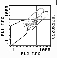
Hello Flowers:

We have started working with JC1 and have noted some interesting
observations.
The setup:
First I should say that we are working with adherent mouse lung cells.
We are pretreating the cultures(in culture) with several compounds that
"reduce" the  mitocondrial membrane potential(MMP). These are:
Valinomycin@10uM for 10 minutes, FCCP@ 2uM for 10 and 30 minutes and
H2O2 @ 300uM for 2hrs. We are also using a positive inducer of MMP,
Gramicidin@10ng/mL for 10 minutes. We are examining the preps thoroughly
under fluorescent microscopy before flow cyto.
I have gathered as many papers as possible on this subject, but
compensation is still a concern of mine. I have a high fl2-fl1, but it
seems to discriminate best here when comparing the fluorescent
microscopy observations.

The observations I have are:

We found compensation to work such that fl2-fl1=50%, while fl1-fl2 is
4.7%.

We are noting a strong increase in JC1 aggregate formation(red) with the
FCCP at 30 minutes and almost no change at the 10 minute exposure when
comparing to happy untreated cells. We expected green depolarized MMP in
both. Why?

The gramicidin increased the red JC1 Aggregate formation, but not
significantly. Why?

Valinomycin seems to work well, however, under epi-microscope there is
red formation amoung the plasma membrane. This affects the flow data.
What is this caused from?

I have attached three jpeg and was hoping that someone could comment on
these.
As we have seen, there doesnt really seem to be an increase in red in
fl2, at any compensation, but rather a decrease in green. We are able to
discriminate at this point but wanted an experienced JC1 user to comment
on this.
The jpeg key is below.

FCCP=red
gramicidin=red+green
H2O2= All green

Scott Tighe
Vermont Cancer Center
Flow Cytometry Core Lab
Burlington, VT


This archive was generated by hypermail 2b29 : Sun Jan 05 2003 - 19:01:16 EST