About Flow Cytometry |
||
|
|
|
||||||
|
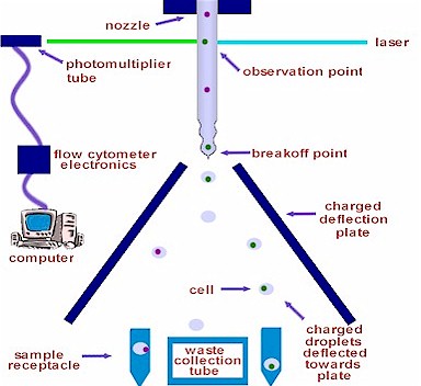
Flow Cytometry DetailsA flow cytometer identifies and verifies events to be analyzed and/or sorted from a sheath flow and droplet stream. Lasers are utilized for excitation and fluorescence of known fluorochromes that have been bound to specific cells or other biological particles. For sorting, once the events are identified, verified and timed in the sheath flow, each event is independently tagged by an electrical charge and passed through oppositely charged deflection plates with ground planes to sort the desired cells from the sample stream.
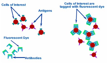
Labeling the Cells of Interest
Cell Analysis
Fluorescence pulses are collected by appropriate optics that focus the light on a sensitive detector, usually a photomultiplier tube (pmt). The detector transforms the pulses of light into electrical pulses that are converted into digital values by the flow cytometer's electronics and then sent to a computer. Each cell also causes scattering of the excitation light. Scattering is a function of the size, shape, and structure and is recorded for each cell. Cell Sorting
The charged droplets pass through a pair of vertical deflection plates, one charged at a negative voltage and the other at a positive voltage. The positively charged droplets shift toward the negative plate and the negatively charged droplets shift toward the positive plate. The droplets then fall into sample receptacles below. Uncharged droplets continue in a straight line out of the flow chamber to an aspirated waste collection tube below. This charging of the droplets allows sorting of particles with two attributes per run by positive or negative charging. The MoFlo cytometer can sort four samples of interest per run by charging the events with a range of electrical charges. |