 |
Achieve stable, uniform, intense, and reproducible
fluorescent labeling of live cells with PKH kits from Sigma.
Sigma is the only provider of PKH Fluorescent Cell Linker Kits for in vivo
or in vitro cell tracking.
|
 PKH Linker Kits for Fluorescent Cell Labeling |
| Named for their discoverer, Paul Karl
Horan, these patented fluorescent dyes and cell labeling technology2 have
been used successfully with animal, plant, and bacterial cells, and even non-cellular
membrane-containing particles.3 Labeled cells can be studied in culture
and in vivo. Rapidly dividing cells like hybridomas, as well as non-proliferative
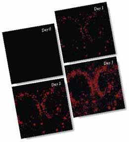
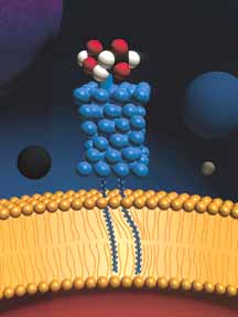
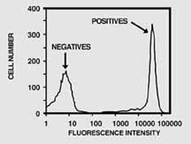
cells like red blood cells, can be labeled and tracked. Easy to use. When you buy from Sigma, you get the PKH dyes, the specially formulated diluents, and the tested labeling protocols all in kit form. Weíve already determined the optimum combinations of reagents and conditions for intense, fast and reproducible viable-cell labeling in many model systems. Our kits make PKH labeling easy. Versatile. The versatility of PKH cell-labeling technology is attributable to its innovative chemistry. Intensely fluorescent dye moieties are attached to long, lipophilic tails. During the short general membrane staining procedure -- only 1-5 min. -- the lipophilic tails diffuse into the cell membrane, leaving the fluorogenic moiety exposed near the outer surface of the cell (Figure 1). Table 2 summarizes some common applications by cell type. Stable labeling without experimental artifacts. The stable partitioning of these molecules into the membrane permits long-term monitoring while leaving the important functional surface proteins unaltered. In fact, all cellular functions, from proliferation to cell-surface antigen recognition, are largely unaffected by PKH dye tagging. In short, labeled cells behave just like unlabeled ones; theyíre just easier to track. Intense, reproducible membrane staining. PKH dye labeling protocols can usually be optimized to give over 75% viable labeled cells with mean fluorescence more than 1,000 times background (see Figure 2). Such high signal-to-noise ratios are especially important when tagging proliferating cell lines because each daughter cell gets only half its parentís label. With PKH dyes, up to ten generations can be followed before the intensity of the label falls near background. Label by phagocytosis. Phagocytic cells can be selectively labeled in the presence of non-phagocytic cells by using an alternate diluent. In this diluent, dyes do not dissolve, but instead form microaggregates; these microaggregates cannot become embedded in membrane, but can be ingested by phagocytosis. This method has been used to study lifetime and migrational patterns of macrophages and neutrophils in vivo. |
Table 2 Some documented applications of PKH dye labeling by cell type. For references to specific applications, call Technical Service. 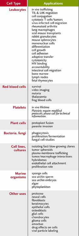 |