Main MenuOn-Line CatalogTechnical LibraryFields of InterestWhat's NewOn-line OrderingSigma Service


PKH Linker Kits

Characteristics of PKH Dyes
Methods for General Cell Labeling
References/Product Information


Characteristics of PKH Dyes
Three different PKH dyes are used for labeling cells: PKH2, PKH26, and PKH67.


PKH2 and PKH67 are green fluorescent labels; both can be detected with microscopes and flow cytometers equipped with fluorescein filters. Since their emission spectra have little overlap with the red region, they are ideal for cytotoxicity studies4,5 in conjunction with red fluorescent viability probes like propidium iodide (PI) and 7-aminoactinomycin D (7-AAD), as well as with other red labels like R-phycoerythrin and Texas Red. They are also excellent for plant studies because they avoid interference from chlorophyll auto-fluorescence in the red region.


The in vivo half-life of PKH2 is 5-8 days, making it excellent for short- to medium-term studies. PKH67 has longer aliphatic tails than PKH2, resulting in more stable labeling and less cell-to-cell transfer.6 It is expected to have an in vivo half-life of 10-12 days. Previous users of PKH2 may find that PKH67 gives even better results for longer-term studies and for studies in which it is essential to minimize cell to cell transfer.


PKH26 is a red fluorescent dye. It offers the longest in vivo half-life -- greater than 100 days -- making it ideal for in vivo cell tracking, cell proliferation studies, and other long-term assays.7-9 Rhodamine or phycoerythrin (PE) filters are suitable for PKH26 detection. Standard fluorescein excitation wavelengths may be used, but fluorescence intensity will be somewhat reduced.


PKH26 is complementary to the green PKH dyes. It can be quantitated without interference from PKH2, PKH67, or fluorescein-tagged antibodies in studies of mixed cell populations. Sigma’s Cell Census PlusTM System builds on the unique properties of PKH26: using flow cytometry, you can measure proliferation and quantitate different phenotypes simultaneously in a mixed cell population.8,10
Figure 1
Schematic of a PKH dye-labeled cell membrane.


Figure 2
Typical fluorescence intensity spectrum of PKH dye-labeled vs. unlabeled cells.


Table 3
Properties of fluorochromes used in cell labeling.

Table 4
Optimum cell and dye concentrations vary with cell type. In your own application, labeling conditions will depend on whether viability or labeling intensity is the most important factor. These combinations have been used successfully in published studies.

Methods for General Cell Labeling
The in vitro general cell labeling procedure can be used for monocytes, macrophages, lymphocytes, and other cells in suspension. Cells grown on the surface of tissue culture vessels may be stained in situ but heterogeneous staining may result. For homogeneous staining, adherent cells should first be suspended with a proteolytic treatment (e.g., trypsin + EDTA). Figure 3 represents the protocol schematically.


A 2x cell suspension and a 2x dye solution, both in the PKH diluent supplied with the kit, are mixed and incubated briefly at room temperature. The labeling reaction is stopped by addition of protein (medium with serum or BSA). Labeled cells are washed 3-5 times to remove unbound dye. General cell labeling should be performed prior to monoclonal antibody staining to avoid capping the antibody with the dye.


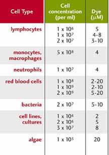
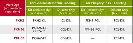
Table 4 gives cell and dye concentrations that have been used successfully for many different cell types. These concentrations may suggest a starting point when you determine the optimum labeling conditions for your cell type and experimental 
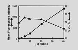
purposes. Optimization is a simple process of determining the dye concentration that gives the best balance of fluorescence intensity and viability (or other measures of cell function).


References

  1. Samlowski, W.E., et al., J. Immunol. Methods, 144, 101 (1991).
  2. The patented PKH dye technology, developed by Zynaxis Cell Science, is now owned by Phanos Technologies.
  3. Horan, P.K., et al., Methods in Cell Biology, 33, 469 (1990).
  4. Slezak, S., and Horan, P., J. Immunol. Meth., 177, 205 (1989).
  5. Hatam, L., et al., Cytometry, 16, 59 (1994).
  6. Unpublished data, Zynaxis, Inc.
  7. Horan, P., and Slezak, S., Nature, 340, 167 (1989).
  8. Yamamura, Y., et al., Cell. Molec. Biol., 41, S121 (1995).
  9. Wallace, P., et al., Cancer Res., 53, 2358 (1993).
  10. Hendrikx, P., et al., Exper. Hematol., 24, 129 (1996).

    Texas Red is a Trademark of Molecular Probes, Inc.

    Cy is a Trademark of Amersham Life Science, Inc.
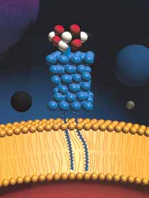
Figure 3
Standard protocol for PKH dye labeling with Sigma kits.
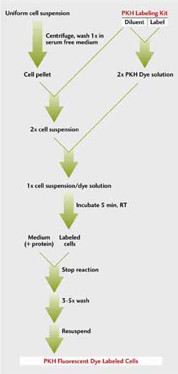
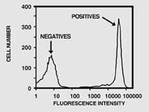


Figure 4
Typical optimization data for PKH dye labeling protocols.9 (Reprinted with permission)


Product numbers of Sigma PKH kits and diluents. The patented diluents are essential components for rapid, uniform, stable labeling.


* CCPS-1, the Cell Census Plus™ System Kit, contains additional components for flow cytometric proliferation studies.

Covered by U.S. Patent Numbers 4,783,401; 4,762,701; and 4,859,584.
Distributed for Phanos Technologies.

Click products in table for data sheets.

For detailed protocols or a complete bibliography, contact your local Sigma Technical Service office or call Immunochemicals Technical Service (800-262-9141 or 314-771-5765 collect).


For laboratory use only. Not for household, drug or other uses.


| Main Menu | Online Catalog | Technical Library | Fields of Interest |
|
What's New | On-line Ordering | Sigma Service | Site Index |

Sigma