Fern spore slide was used in our experiment. A
Nikon 40x NA 1.30 oil-immersion objective is used for imaging. The Z-axis step is set
to 1μm per image, and 60 successive images are collected. The driving
voltage is
±3V
for both galvanometers. The depth-resolved diffused reflection images are
shown in the animation in Fig. 1. For comparison, a
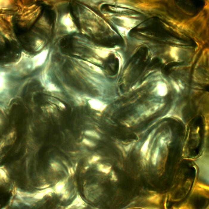
QImaging Retiga 1300i 12-bit
digital color CCD camera is used to get the image of the same spore with a
Nikon 1x relay lens. The wide-field color image is shown in Fig. 2. After
the collection of the depth-resolved image, 3-D projection is performed with
ImageJ as shown in Fig. 3. As can be seen from the animation that, the 3-D
projection result of the spore clearly indicates the spatial structure and
position of each prothallium. Fig. 4
shows the corresponding results obtained from the Bio-rad MRC-1024 confocal
system with the same objective. It can be seen that, the results obtained
from our CSLM system is comparable to the commercial ones.