|
Overview
This project chronicles handmade and mould made paper images produced
by a scanning-laser confocal microscope. This type of microscopy is
unsurpassed for producing sharp three-dimensional images and allows
us to see not just surface features, but often all the way through
the sheet. The microscope scans successively deeper layers of a specimen
and then uses a computer to assemble the various images into a single
composition. This technique eliminates blurring and scatter. The resulting
compositions have proved to be not just helpful in understanding how
the fibers are interlaced within a specific paper, but are also esthetically
of interest as works of art.
All about the Paper Project People
Charles Kazilek has
been a member of the School of Life Sciences at Arizona State University
(USA) since 1985. He is currently the manager of the Life Science
Visualization Group and the Technical Director of the William M. Keck
Bioimaging Laboratory. He teaches classes in scientific data presentation
and advanced bioimaging. His daily scientific work is complemented
by his fine arts activities as a painter and photographer
Gene Valentine is Professor Emeritus of English at Arizona State
University and proprietor (since 1979) of the Almond Tree Press and
Paper Mill in Tempe, Arizona. His interest in the interior structure
of his own silk and cellulose fiber papers led to his current collaboration
with Charles Kazilek and their work with the Leica scanning-laser
confocal microscope at Arizona State University.
Confocal images were produced in the William M. Keck Bioimaging
Laboratory, Arizona State University.
3-D images
Some of the images in our galleries can be viewed in three dimensions.
This requires a pair of red-blue 3-D glasses also called anaglyph
glasses.
 |
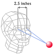
With two eyes we have the ability to see depth,
or as it is sometimes called 3-D. Depth occurs when images from
two different angles are interpreted by our brain.
Our eyes are separated by a distance of about 2.5 inches,
and because they are separated, we see objects from two different
angles. |
3-D images mimic what our eyes do naturally. An anaglyphic 3-D picture
is made from two different images. In most cases, two pictures are
taken with a camera. Each image is taken at a slightly different angle
similar to the angle each of our eyes normally sends to the brain.
The left image is colored red and the right image is colored blue.
The images are combined into one picture for 3-D viewing. At this
point the 3-D picture looks blurred to the naked eye.
When you put on 3-D glasses with red and blue filters, your left
eye sees only the red image and your right eyes sees only the blue
image. Your brain combines the images from each eye to "see" depth
or 3-D.
For the Paper Project 3-D images, two different projections were
made using a computer. Projections can be made at slightly different
angles so they mimic the angle each eye would naturally see. The projections
are colored red for the left eye and blue for the right eye. The images
are combined, and viewed using 3-D glasses. Thanks to our brain, the
combined images now show the fibers of paper in 3-D.
What does the word 'anaglyph' mean?
an-a-glyph (an'-a-glif). - an image made up of two slightly different
views (angles), in contrasting colors, of the same subject; when viewed
using a pair of corresponding filters, the picture appears three-dimensional.
|