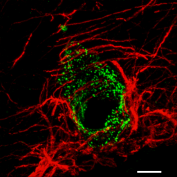
Image of the blood-brain
barrier characterized by microvascular cerebral endothelial
cells showing Factor VIII positivity (pseudo-coloured
green) surrounded by astrocytic foot processes (pseudo-coloured
red). The image is a maximum-intensity projection of
a confocal z-series. Sample courtesy of Dr. S. Nag
and R. Venugopalan (TWRI). Scale bar 10 μm.
For details see: Nag,
S. (2003). Immunohistochemical detection of endothelial
proteins. Methods Mol. Med. 89:489-501.
|