|
A.O. Brightman1, B.P. Rajwa2, J.E. Sturgis2, M.E. McCallister1, J.P. Robinson2,
1Dept. of Biomedical Engineering,
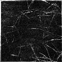
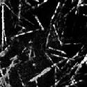
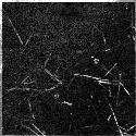
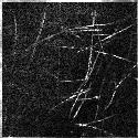
Time-Lapse Reflection Imaging of Matrix Assembly - Collagen, type I, or pepsin-solubilized intestinal submucosal, PSIS, was polymerized in 8-well chambered coverglass (Lab-Tek, Nalge Nunc Int.). The unstained 3-dimensional matrices were viewed with an inverted confocal laser-scanning microscope (BioRad MRC1024) using a 60X, oil immersion lens. Laser light of 488 nm illuminated the hydrated samples and the reflected light was detected with a photomultiplier tube using a blue reflection filter. Samples were maintained at 30-37°C on a heated stage. Time-lapse images of fibrillogenesis were recorded at 10-15 second intervals. For standardizing the scanning depth in the matrix, the Z position was kept in the range of 20 to 50 µm from the coverglass.
All material (text, images and movies) copyright Purdue University. No material may be reproduced in any form without the express permission of Purdue University. |
Video 1 (2.5 MB, 17 sec) PSIS assembly real time: 60 min 
|