| The Phosphatidyl Serine Detection Kit | ||
The Phosphatidyl Serine Detection™ Kit to measure apoptosis: The significance of apoptosis in clinical research is just now beginning to be appreciated. Apoptosis or programmed cell death (PCD) is a genetically encoded cell elimination program which ensures an equilibrium between cell proliferation and cell death and by which damaged or unwanted cells are eliminated. Therefore it is important to stress that apoptosis is a normal physiological process. Without continuous signalling by growth factors, hormones or cytokines, all cells undergo PCD. Aberrations in the mechanism of apoptosis occur in congenital defects, malignancies, auto-immune diseases, immune deficiency syndromes and in degenerative conditions (1,2).
The Phosphatidyl Serine detection kit provides a rapid and reliable method for the detection of
apoptosis by flow cytometry. The method enables detection at the single-cell level (3) and also
allows the distinction between apoptosis and necrosis. During the early stages of apoptosis,
phosphatidyl serine (PS) becomes exposed on the outside of the cell membrane. In vivo, this is a
signal to phagocytes to engulf the dying cell before it looses its plasma membrane integrity and
releases inflammatory mediators into the surroundings. This early stage of apoptosis can be
specifically detected by PS binding proteins, such as Annexin V. During the early stage of apoptosis,
the cell membrane is intact and the cells exclude propidium iodide (PI). Later, during in the
apoptotic process in vitro, the membrane becomes porous and PI becomes associated with DNA in the
nucleus. The uptake of PI is an indication of necrosis. In a single cell suspension, these events can
be analysed using the Phosphatidyl Serine detection kit, which contains the PS binding protein,
Annexin V FITC or PE, plus calcium buffer and PI. (See table 1 below). The binding of Annexin V to
phosphatidyl serine during early apoptosis is illustrated in Figure 1.
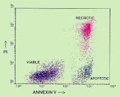 Figure 1. Flow cytometry analysis of peripheral blood lymphocytes labelled with Annexin V FITC and PI after overnight incubation with dexamethasone. Apoptotic cells exclude PI and express phosphatidyl serine which is detected by Annexin V FITC. Necrotic or dead cells are permeable for PI which associates with nuclear DNA and is visible as red fluorescence. The dot plot shows three populations: viable cells (lower left), apoptotic cells (lower right) and apoptotic cells which have lost plasma membrane integrity (upper right).
|
 |
    |
 |
|---|---|---|
| ||