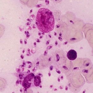
 Plasmodium falciparum malaria trophozoites and gametocyte.
Plasmodium falciparum malaria trophozoites and gametocyte.
Plasmodium falciparum malaria trophozoites and gametocyte.
 Plasmodium falciparum - several stages
 An EM image of an infected red cell.
 Autofluorescing C.E. porphyria red cells.
 Sunlight induced lesions on the hand of this young child.
 Kala Azar (visceral Leishmaniasis) in a bone marrow macrophage.
Back to clinical immunophenotyping
 |
 |
 |
 |
 |
CD-ROM Vol 3 was produced by Monica M. Shively and other staff at the Purdue University Cytometry Laboratories and distributed free of charge as an educational service to the cytometry community. If you have any comments please direct them to Dr. J. Paul Robinson, Professor & Director, PUCL, Purdue University, West Lafayette, IN 47907. Phone:(765) 494-0757; FAX (765) 494-0517; Web http://www.cyto.purdue.edu, EMAIL cdrom3@flowcyt.cyto.purdue.edu