Apoptosis is a cell death process characterized by morphological and
biochemical features occurring at different stages. Once triggered, apoptosis
proceeds with different kinetics depending on cell types and culminates
with cell disruption and formation of apoptotic bodies. A critical stage
of apoptosis involves the acquisition of surface changes by dying cells
that eventually results in the recognition and the uptake of these cells
by phagocytes. Different changes on the surface of apoptotic cells such
as the expression of thrombospondin binding sites, loss of sialic acid
residues and exposure of a phospholipid-like phosphatidylserine (PS) were
previously described. Phospholipids are asymmmetrically distributed between
inner and outer leaflets of the plasma membrane with phosphatidylcholine
and sphingomyelin exposed on the external leaflet of the lipid bilayer,
and phosphatidylserine predominantly observed on the inner surface facing
the cytosol.
Exposure of PS on the external surface of the cell membrane has been
reported for activated platelets and senescent erythrocytes. Recently,
it was shown that cells undergoing apoptosis break up the phospholipid
asymmetry of their plasma membrane and expose PS which is translocated
to the outer layer of the membrane. This occurs in the early phases of
apoptotic cell death during which the cell membrane remains intact. This
PS exposure may represent a hallmark (early and widespread) in detecting
dying cells. Annexin V, belonging to a recently discovered family of proteins,
the annexins, with anticoagulant properties has proven to be a useful tool
in detecting apoptotic cells since it preferentially binds to negatively
charged phospholipids like PS in the presence of Ca2+ and shows minimal
binding to phosphatidylcholine and sphingomyeline. Changes in PS asymmetry,
which is analyzed by measuring Annexin V binding to the cell membrane,
were detected before morphological changes associated with apoptosis have
occurred and before membrane integrity has been lost. By conjugating FITC
to Annexin V it is possible to identify and quantitate apoptotic cells
on a single-cell basis by flow cytometry. Staining cells simultaneously
with FITC-Annexin V (green fluorescence) and the non-vital dye propidium
iodide (red fluorescence) allows (bivariate analysis) the discrimination
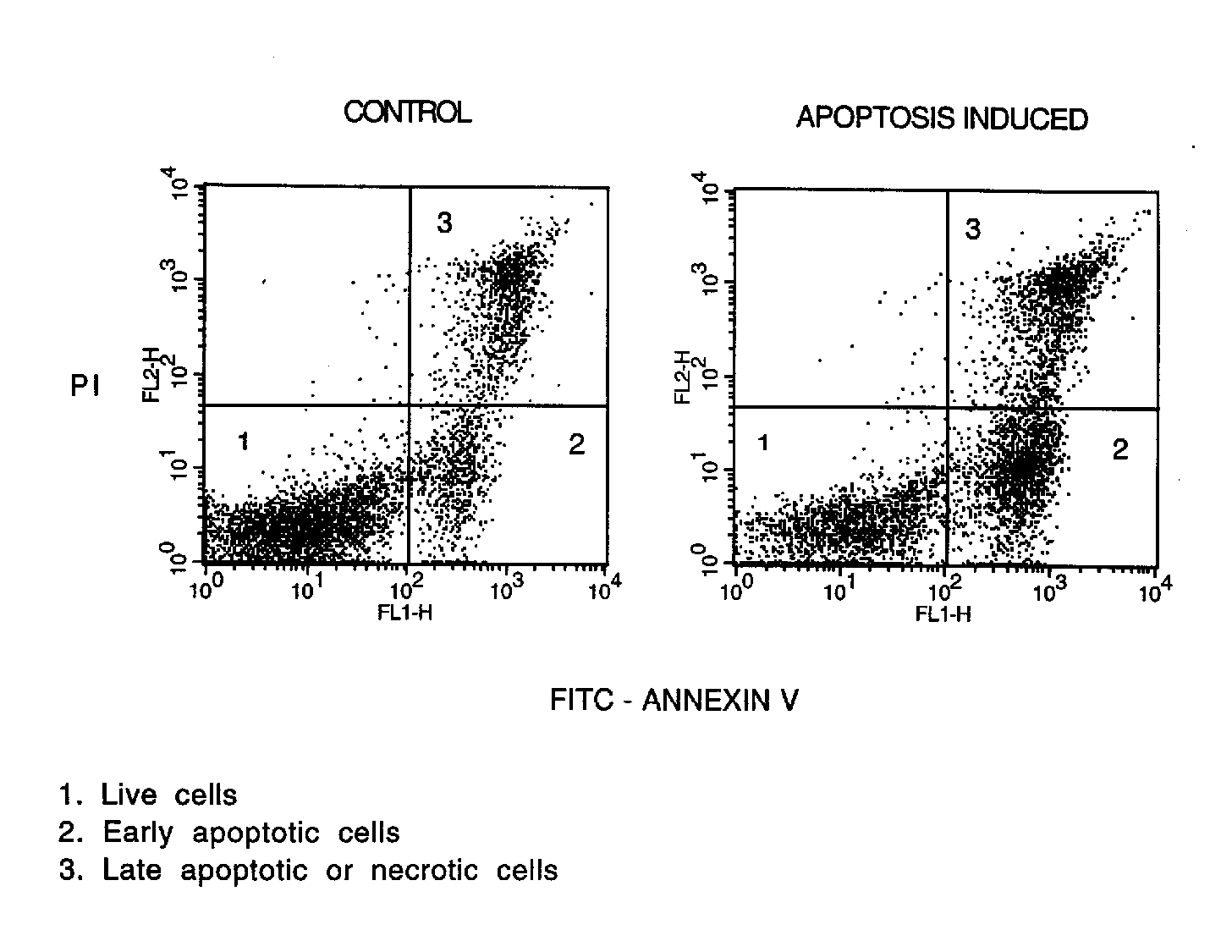
of intact cells (FITC-PI-),
early apoptotic (FITC+PI-) and late apoptotic or necrotic cells (FITC+PI+).
2A. Basic protocol
2A.1. Materials
FITC-labeled recombinant Annexin V (A2)
Binding buffer (A1)
Propidium iodide (PI) solution (A1)
Cytofluorimeter equipped with an argon laser with excitation at 488
nm (A3)
2A.2. Methodology1. Wash 2x 1x106 cells with PBS.
2. Dilute FITC-Annexin V at a concentration of 1 mg/ml in binding buffer
and resuspend cells in 1 ml of this solution (prepare it freshly each time).
3. Incubate 10 min in the dark at RT.
4. Add to the cell suspension 0.1 ml of PI solution prior to analysis
to give a final concentration of 1 mg/ml.
5. Analyze cells by flow cytometry:
- acquire data with CellQuest or LYSYS II or any software for phenotyping
analysis;
- collect 10,000 events per sample;
- exclude debris by scatter gating (forward vs. side);
- display data as two-color dot plot with FITC-Annexin V (green fluorescence,
X axis) vs. PI (red fluorescence, Y axis).
2B. Support Protocols
2B.1. To detect apoptosis and cell phenotype at the same time,
cells can be first incubated with the appropriate predetermined concentration
of PE-conjugated mAb, then washed with PBS and incubated with FITC-Annexin
V according the basic protocol. The cytofluorimetric analysis is performed
with PE (orange) as third fluorescence.
2B.2. Annexin V can also be conjugated to biotin. The secondary
detection reagent can be the streptavidin-FITC conjugate. To this purpose
follow the basic protocol except for substituting FITC-AnnexinV with biotin-Annexin V, then wash cell with binding buffer; add the secondary reagent and incubate 20 min at RT. Continue with the basic protocol from step 4.
3. Commentary
3.1. Background information
Loss of plasma membrane asymmetry seems to be a universal phenomenon
of apoptosis, thus all cell types undergoing apoptosis can be quantitated
by staining with Annexin V-FITC and PI. Apoptotic cells become Annexin
V positive after nuclear condensation has started, but before the cell
becomes permeable to PI. This binding assay compared with traditional methods
like microscopy, DNA electrophoresis, DNA flow cytometry appears to be
sensitive, it correlates with other tests, and it is easy, fast and reliable
to perform. Different from other methods which measure cells that have
already reached the stage where a substantial amount of the DNA has been
fragmented and leaked from the cell or has been altered and lost staining
capacity, the Annexin V binding test allows the quantitation of cells at
early stages of apoptosis or when apoptosis can occur in absence of DNA
fragmentation, the discrimination between apoptosis and necrosis, and the
simultaneously identification of the cell surface markers. The Annexin
V binding method was established with peripheral lymphocytes and neutrophils,
germinal centre B cells, rat thymocytes and Burkitt's lymphoma cell lines.
It should be suitable for any cell type growing in suspension (for adherent
cells see 3.2: Critical Parameters). This methodology can also be used
to detect apoptotic cells under a microscope.
3.2. Critical parameters
This technique is not suitable for cells from solid tissue or adherent
cells because of the damage to plasma membrane caused in the attempt to
disaggregate cells to have a single cell suspension. Any procedure which
affects the integrity of the plasma membrane will result in cell positive
for Annexin V. PI stainability provides evidence of the loss of plasma
membrane integrity. The binding of Annexin V to phosphatidylserine can
be affected in adherent cells, which are usally detached from plastic dishes
by enzymatic treatment, although their membrane integrity is not altered.
Care should be used in preparatory procedures: true apoptotic cells should
be those cells which exclude PI and exhibit phosphatidylserine on the outside
of the plasma membrane.
3.3. Troubleshooting
a) Absence of FITC-Annexin V fluorescence: apoptosis
was not induced in the cells; inappropriate dilution of FITC-Annexin V
reagent. Use as control a well documented stimulus and cell to induce apoptosis.
Check for the right FITC-Annexin V dilution.
b) Elevated FITC-Annexin V and/or PI stainability: apoptosis is
an ongoing process so that cells stained with Annexin V should not be kept
for prolonged times before measurement. Cells which still mantain membrane
integrity for longer incubation times may become positive for PI since
this dye will enter intact cells although very slowly. Analyze cells as
soon as are stained and add PI solution just before the analysis.
3.4. Anticipated results
The biparametric analysis of FITC-Annexin V green fluorescence (X axis)
versus PI red fluorescence (Y axis) of control cells should represent a
cytogram in which the majority of cells remains alive showing only a background
level of PI and Annexin V staining confined to the lower left quadrant.
The cytogram of cells undergoing apoptosis should show the early apoptotic
cells in the lower right quadrant being Annexin V positive and PI negative;
late apoptotic or necrotic cells are in the upper right quadrant being
PI positive and Annexin V positive; live cells in the lower left quadrant
being negative for both fluorescent probes (Figure 1).
3.5. Time considerations
The actual time required to perform this method is quite short being
about 15 min for the cell staining and 10 min for analysis (depending on
the number of samples). Variable times are required to prepare cells to
be stained .
3.6. Key references
1. Koopman, G., Reutelingsperger, C. P., Kuijten, G. A. M., Keehnen,
R. M. J., Pals, S. T., and van Oers, M. H. J. 1994. Annexin V for flow
cytometric detection of phosphatidylserine expression on B cells undergoing
apoptosis. Blood 84: 1415.
2. Homburg, C. H., de Haas, M., von dem Borne, A. E., Verhoeven, A.
J., Reutelingsperger, C. P., and Roos, D. 1995. Human neutrophils lose
their surface Fc gamma RIII and acquire Annexin V binding sites during
apoptosis in vitro. Blood 85: 532.
3. Vermes, I., Haanen, C., Steffens-Nakken, H., and Reutelingsperger,
C. 1995. A novel assay for apoptosis - flow cytometric detection of phosphatidylserine
expression on early apoptotic cells using fluorescein labelled Annexin
V. J. Immunol. Meth. 184: 39.
4. Fadok, V. A.,Voelker, D. R., Campbell, P. A., Cohen, J. J., Bratton,
D. L., and Henson, P. M. 1992. Exposure of phosphatidylserine on the surface
of apoptotic lymphocytes triggers specific recognition and removal by macrophages.
J. Immunol. 148: 2207.
Appendix 1 (A1): Stock solutions
Solution
Preparation
Storage
Binding buffer
Hepes buffer: 10 mM HEPES/NaOH, pH 7.4, 150
mM NaCl, 5 mM KCl, 1 mM MgCl2, 1.8 mM CaCl2
RT
PI solution
Propidium iodide 10 mg/ml in binding buffer
RT
Appendix 2 (A2): Reagents
Annexin V-FITC
Alexis 209-250-T010 Bender MedSystem, Boehringer Ingelheim
BMS306FI Boehringer Mannheim1-828-681 Chemicon AG606 Oncogene Research Products PF032 R&D System KNX50