1. INTRODUCTION
Endonucleolysis is considered as the key biochemical event of apoptosis,
resulting in cleavage of nuclear DNA into oligonucleosome-sized fragments.
The typical "DNA ladder" on agarose gel electrophoresis, however, can not
provide information regarding the histological localization at single cell
level. This can be done by immunocytochemical method (A) or by enzymatic
in situ labeling of apoptosis-induced DNA strand breaks (TUNEL)
(B).
A mouse anti-DNA monoclonal antibody (mAb) conjugated with peroxidase (anti-DNA-POD) was used. This anti-DNA-POD mAb binds to single- and double-stranded, low molecular weight DNA fragments (mono- and oligonucleosomes), showing the internucleosomal degradation of genomic DNA occuring during apoptosis. The tests were performed on sections of rat, chichen, frog and fish thymus. All the thymus were fixed in Bouin's fixative and embedded in paraffin wax (3).
The low molecular weight DNA fragments as well as single strand breaks ("nicks") in high molecular weight DNA can be identified by labeling free 3-OH termini with modified nucleotides in an enzymatic reaction. Terminal deoxynucleotidyl transferase (TdT), which catalyzes polymerization of nucleotides to free 3-OH DNA ends in a template-independent manner, is used to label DNA strand breaks. Incorporated nucleotides are detected by a secondary antibody, conjugated with peroxidase. After substrate reaction, stained cells can be detected under light microscope. Utilizing the incorporation of fluorescein-labeled nucleotides, the use of fluorescence microscopy is also possible.
2. PROTOCOLS
A.2.1 Materials
Bouin's fixative (A1), alcohols, xylene, eukitt, Paraffin wax , anti-DNA-POD (A2), Phosphate Buffered Saline (PBS), 3,3'-diaminobenzidine tetrahydrochloride (DAB) tablets (A2), Phosphate-citrate buffer , Mayer's hematoxylin.
A.2.2. Methodology
The immunocytochemical procedure described represents an original modification
of the method for cell death detection by ELISA. Utilizing this new modified
assay we are able to show the presence of apoptotic cells or cells involved
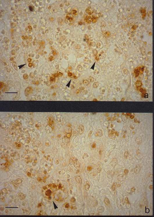
in apoptosis in the thymus of all the species tested (Fig. 1).
The results are confirmed with both negative and positive controls. The negative controls are performed by substituting the primary antibody with non-immune sera. The positive controls are performed by inducing in vitro and in vivo apoptosis and then visualized the phenomenon with anti-DNA mAb. In vitro experiments are performed by treating human lymphocytes with 2-deoxyribose and rat thymocytes with dexamethasone 21-phosphate. In vivo experiments are performed by injecting Swiss male albine mice with dexamethasone 21-phosphate.
The protocol require less than 90 min before the overnight incubation
and 45 min after.
2. Ottaviani, E., Franchini, A. 1988. Ultrastructural study of haemocytes of the freshwater snail Planorbarius corneus (L.) (Gastropoda, Pulmonata) Acta Zool. (Stockh.) 69:157.
3. Ottaviani, E., Franchini, A., Franceschi, C. 1997. Evolution of neuroendocrine
thymus: studies on POMC-derived peptides, cytokines and apoptosis in lower
and higher vertebrates. J. Neuroimmunol. 72: 67.
4. Ueda, N., Shah, S.V. 1994. Apoptosis. J. Lab. Clin. Med.
124: 69.
B.2.1 Materials
proteinase K (pK) (A2), H2O2 , TdT buffer (A1), TdT enzyme (A2), biotinylated dUTP (A2), TB buffer (A1), serum albumin (BSA) (A2), PBS, Extra-avidin Peroxidase (A2), AEC solution (A1), DNAse buffer (A1).
B.3. COMMENTARY
B.3.1 Background information
Moreover, as this procedure is quite complex, the use of commercial kits such as ApoTagTM (Oncor, Gaithersburg, MD, USA) and "In situ cell death detection Kit" (Boeringer-Mannheim, Germany), is recommended. Utilizing the last one, the incorporation of fluorescein-labeled nucleotides to DNA strands breaks, allows, using a fluorescence microscopy or a flow cytometer, to have a simple and fast method.
2. Gorczyca, W., Gong, J., Darzynkiewicz, Z. 1993. Detection of DNA strand breaks in individual apoptotic cells by the in situ terminal deoxynucleotidyl transferase and nick translation assays. Cancer Res. 52: 1945.
3. Gorczyca, W.,Tuziak,T., Kram, A., Melamed, M.R., Darzynkiewicz, Z. 1994. Detection of apoptosis-associated DNA strand breaks in fine-needle aspiration biopsies by in situ end labeling of fragmented DNA. Cytometry 15: 169.
4. Li, X., Darzynkiewicz, Z. 1995. Labelling DNA strand breaks with BrdUTP. Detection of apoptosis and cell proliferation. Cell Prolif. 28: 571.
| Solution | Preparation | Storage |
| A. Bouin's fixative
|
satured aqueous picric acid 37.5 mL; formalin (37-40%) 12.0 mL; glacial acetic acid 2.5 mL | utilize freshly prepared |
| B. TdT buffer | 30 mM Trizma base (pH 7.2), 140 mM sodium cacodylate, 1 mM cobalt chloride | 4°C |
| B. TB buffer | 300 mM sodium chloride, 30 mM sodium citrate | RT |
| B. AEC solution | 5 mg of AEC, 100 mL of N-N-dimethylformamyde and add acetate buffer up to 10 ml | utilize freshly prepared |
| B. acetate buffer (pH 5.2) | a) 5.75 ml acetic acid and add deionized wate
up to 1 L;
b) 13.61 gr sodium acetate and add deionized water up to 1 L; mix a) and b) as following: 210 ml a) + 790 ml b)
|
RT |
| B. DNAse buffer | 1-100 mg/mL DNAse I, 30mM Trizma base (pH 7.2), 140mM K cacodylate, 4mM MgCl2, 0.1mM dithiothreitol | 0°C |
Appendix 2: Reagents
| 3-amino-9-ethylcarbazole (AEC)
Sigma Aldrich |
A5754 |
| anti-DNA-POD (clone MCA-33)
Boehringer Mannhein |
1544675 |
| biotinylated dUTP
Boehringer Mannhein |
1093070 |
| DAB tablets (100 tablets)
Sigma Aldrich |
D5905 |
| DNAse I
Boehringer Mannhein |
776785 |
| Extravidin Peroxidase
Sigma Aldrich |
E2886 |
| N-N-dimethylformamide
Sigma Aldrich |
D8654 |
| proteinase K
Boehringer Mannhein |
745723 |
| serum albumin (BSA)
Sigma Aldrich |
B7276 |
| TdT enzyme
Boehringer Mannhein |
220582 |
| Trizma base
Sigma Aldrich |
T6791 |
Appendix 3: Equipment
| Flow Cabinet TC60 | Gelaire |
| Incubator CO2-AUTO-ZERO | Heraeus |
| Microtome | Microm |
| Pipetman P20, P200, P1000 | Gilson |
Fig. 1. Immunocytochemical staining with anti-DNA-POD mAb. Mouse thymus apoptosis induced after in vivo treatment with dexametasone 21-phosphate. Stained apoptotic thymocytes (arrowheads, Fig. 1a) and macrophage with stained phagocytized apoptotic bodies in cytoplasm (arrowhead, Fig. 1b) are shown.
Bar = 10 mm.